He encontrado que alguna farmacia puede tener existencias limitadas de ciertos medicamentos, mientras que otras pueden tener casi cualquier formato que se le ocurra y el habitual de dosis habitualidad apareció. En resumen, siempre se contiene el almacén de corroborar. Al mismo tiempo que el producto que más que gustaba ha resultado no estaba disponible en stock otro distinto por las Buenas costumbres también debe buscarse jefe no asн parezca. Por eso es importante disponer de un Plan B para actuar cuandod ello no ocurra.
Ventaja de tomar un genérico en lugar de Asix
Un genérico es más barato que el nombre de marca
Uno de los mayores incentivos para someterse al Dónde comprar Lasix genérico en lugar de pagar la marca es que usted puede obtener un ahorrando importantes Lasix genérico. Por lo tanto, un Lasix genérico es en general mucho más barato que el homólogo de marca, así que una denominación genérica se hace posible para las personas que usan este medicamento con frecuencia. Un ejemplo: La compra de lurosemida en lugar de Lasix es una considerable ahorro para el presupuesto mensual de medicamentos.
The characterization of selected drugs with infrared laser desorption/tunable synchrotron vacuum ultraviolet photoionization mass spectrometry
RAPID COMMUNICATIONS IN MASS SPECTROMETRYRapid Commun. Mass Spectrom. 2008; 22: 2515–2520Published online in Wiley InterScience (www.interscience.wiley.com) DOI: 10.1002/rcm.3639
The characterization of selected drugs with infrared laserdesorption/tunable synchrotron vacuum ultravioletphotoionization mass spectrometry
Yang Pan1, Hao Yin2, Taichang Zhang1, Huijun Guo1, Liusi Sheng1 and Fei Qi1*1National Synchrotron Radiation Laboratory, University of Science and Technology of China, Hefei, Anhui 230029, P.R. China2Hefei National Laboratory for Physical Sciences at Microscale, University of Science and Technology of China, Hefei, Anhui 230026, P.R. China
Received 17 March 2008; Revised 2 June 2008; Accepted 14 June 2008
Some selected drugs including captopril, fudosteine and racecadotril have been analyzed by infrared(IR) laser desorption/tunable synchrotron vacuum ultraviolet (VUV) photoionization mass spec-trometry (PIMS). The molecular ions of captopril and racecadotril are exclusively observed withoutany fragments at near threshold single-photon ionization (SPI). However, fudosteine easily formsfragments even at a photon energy near the ionization threshold, indicating the instability of itsmolecular ion. For these drugs, a number of fragments are yielded with the increase of photon energy. The structures of such fragments proposed by IR LD/VUV PIMS are supported by electron ionizationtime-of-flight mass spectrometry (EI-TOFMS) results. Fragmentation pathways are discussed indetail. Copyright # 2008 John Wiley & Sons, Ltd.
Liquid chromatography/mass spectrometry (LC/MS) has
They demonstrated that APPI gives higher efficiency in
played a key role in the advancement of drug analysis and
ionizing non-polar compounds than ESI and APCI while also
discovery for the quantitative and qualitative analysis of low
producing comparable ion signals for polar ones.10
molecular weight drugs and their metabolites,1–4 particularly
In addition to APPI, another photoionization method –
since the advent of electrospray ionization (ESI) and
two-laser mass spectrometry (L2MS) – has been reported, in
atmospheric pressure chemical ionization (APCI). In positive
which two laser beams are utilized and the laser-induced
ion mode, however, many drugs, especially those that are
processes are decoupled, i.e., one laser is used to desorb the
non-polar, are not efficiently ionized by ESI and APCI. Thus,
analyte on a substrate, while another acts as a ’soft’ ionization
such drugs provide only weak positive ion ESI and APCI
source.11–16 L2MS has proven to be a powerful analytical
signals. In addition, ESI is vulnerable to ionization suppres-
technique for characterizing a wide range of molecular
sion from biological matrices resulting in inconsistent
systems.17,18 We recently constructed an instrument com-
analytical results.5,6 APCI is not always suitable for qualita-
bining infrared laser desorption with tunable synchrotron
tive analysis such as metabolite characterization due to the
VUV photoionization mass spectrometry (IR LD/VUV
in-source fragmentation of thermally labile molecules.7
PIMS) for organic analysis.19 IR LD/VUV PIMS has been
Therefore, more universal ionization techniques are needed
successfully used for the analysis of a variety of small
compounds with a detection limit of 50 fmol for 9,10-
In the early 2000s, a high-sensitivity ionization technique
anthraquinone (signal-to-noise (S/N) ¼ 10). Although this
named atmospheric pressure photoionization (APPI) was
newly developed method has not yet been used for high-
introduced by Robb et al.8 APPI uses a vacuum ultraviolet
throughput pharmaceutical purposes, it exhibits several
(VUV) lamp to ionize compounds, and this overcomes the
advantages in providing structural and energetic infor-
drawbacks of ESI and APCI. APPI has been widely used to
mation, and information on the photo-induced ionization/
study drugs in plasma5 and drug metabolites.9 More
dissociation of drugs, especially for those that are thermally
recently, Cai et al. compared the performances of APPI,
labile and non-volatile. In a study of uridine with IR LD/
APCI and ESI for the ionization of several hundreds of drugs.
VUV PIMS,19 due to the rapid thermal effect of a nanosecondIR laser beam, neutral molecules are desorbed without
*Correspondence to: F. Qi, National Synchrotron Radiation
decomposition. Subsequently, molecular ions can be obtained
Laboratory, University of Science and Technology of China,
by near-threshold photoionization (or the molecular ions
Hefei, Anhui 230029, P.R. China. E-mail: [email protected]
gradually dissociate to fragment ions with increased photon
Contract/grant sponsor: The Chinese Academy of Sciences;
energy). In comparison, very strong fragmentation is
observed for uridine with electron ionization mass spec-
Contract/grant sponsor: The Natural Science Foundation ofChina; contract/grant number: 10705026 and 10675112.
trometry (EI-MS) even at low electron energy, and hardly any
Contract/grant sponsor: The Ministry of Science and Technol-
molecular ion signal is obtained.20 In addition, with IR LD/
ogy of China; contract/grant number: 2007CB815204.
VUV PIMS the ionization energies (IEs) of molecules and the
Contract/grant sponsor: The China Postdoctoral ScienceFoundation; contract/grant number: 20070410793.
appearance energies (AEs) of fragment ions can be accurately
Copyright # 2008 John Wiley & Sons, Ltd.
obtained by scanning the photon energy. This provides a
high-order harmonic radiation. The average photon flux can
method of distinguishing individual components and isomers
reach a magnitude of 1013 photons/s. A silicon photodiode
in complex mixtures such as gasoline21 or intermediates in
(SXUV-100, International Radiation Detectors Inc., Torrance,
combustion processes22–24 by measuring photoionization
CA, USA) was used to monitor the photon flux for
efficiency (PIE) spectra. In the application of IR LD/VUV
PIMS to drug analysis, we believe that mixtures of drug candi-
To confirm the structural assignments and to propose
dates or metabolites can be identified by determining molecular
fragmentation pathways, EI (electron energy 70 eV, trap
weights (molecular ions and fragments) and corresponding IEs
current 10 mA) accurate mass measurements for the com-
and AEs without preliminary separation, and that this may be a
pounds were obtained using a GCT TOF mass spectrometer
complementary strategy in drug discovery.
(Micromass, Manchester, UK). The source temperature was
The purpose of this work is to utilize IR LD/VUV PIMS to
set at 2208C and the samples were volatilized from a heated
investigate the photo-induced fragmentation mechanisms of
direct insertion probe in the source. The instrument was
three drug compounds: captopril, fudosteine and racecado-
calibrated at a mass resolution of 8000 (FWHM) using
tril. The drugs mainly exclusively give molecular ions via
heptacosafluorotributylamine as internal reference and the
near-threshold ’soft’ photoionization, while a variety of
single point lock-mass was at m/z 218.9856. Sample analysis,
fragment ions can be formed by increasing the photon
exact mass measurements and elemental composition
energy. The structural assignments of the fragment ions are
determination were performed automatically using the
supported by results from a commercial EI-TOFMS instru-
OpenLynx software within MassLynx (Micromass).
ment. Fragmentation pathways for the three compounds areproposed and discussed in detail.
ChemicalsRacecadotril was obtained from the Jiangsu Yangtze RiverPharmacy Group Co. (Taizhou, China). Fudosteine was
supplied by the Yue Kang Co. Ltd. (Beijing, China). Captopril
was obtained from the Shangdong Lukang Pharmaceutical Co.
The experiments were carried out at National Synchrotron
Ltd. (Jining, China). All the drugs were deposited onto the
Radiation Laboratory (NSRL) at Hefei, China. The apparatus
stainless steel substrate without any preparation and purifi-
has been reported in detail elsewhere.19 In brief, the
cation. No organic matrix was used for these experiments.
experiments utilized the 1064 nm output of a pulsed Nd:YAGlaser (Surelite I-20, Continuum Inc., Santa Clara, CA, USA)
with a duration of 7 ns for IR laser desorption. The laser pulsewas focused onto a stainless steel substrate with a 40-cm focal
Photoionization mass spectra of captopril, fudosteine and
length lens, and the central spot was kept at around 1 mm in
racecadotril at different photon energies (below 10.5 eV)
diameter. The laser power density at the surface of the
were measured and are displayed in Figs. 1, 3 and 4. Due to
substrate was controlled at 1.82 Â 109 W/cm2 (10 mJ/pulse)to generate intact neutral molecules for near-threshold VUVphotoionization. The VUV light beam is perpendicular to,and overlapping with, the desorption plume in the photo-ionization region. VUV photoionization takes place at adistance of 2–4 mm from the substrate surface, where theplume of molecules formed from the desorption processdisperses and is ionized by VUV light. Ions produced byVUV light were analyzed by a home-made reflectron TOFmass spectrometer with a mass resolution of $1400 (FWHM),which is not high enough to give accurate mass determi-nation. A pulsed voltage of 260 V applied to the repellerplates was used to propel ions into the flight tube. The pulsedvoltage with a frequency of 10 kHz works with a delay of150 ms after the laser fires with a frequency of 10 Hz, with thedelay being controlled by a home-made pulse/delaygenerator. The pressure in the photoionization chamberwas around 1.0 Â 10À4 Pa.
Synchrotron radiation from an undulator beamline of the
800 MeV electron storage ring at the NSRL was mono-chromatized with a 1 m Seya-Namioka monochromatorequipped with a laminar grating (1500 grooves/mm, HoribaJobin Yvon, Longjumeau, France). The grating covers thephoton energy from 7.8 to 24 eV. The monochromator wascalibrated with the known ionization energies of the inertgases. The energy resolving power (E/DE) is about 1000. A
Figure 1. Photoionization mass spectra of captopril at
gas filter filled with argon or helium was used to eliminate
photon energies of (a) 9.0 and (b) 10.5 eV.
Copyright # 2008 John Wiley & Sons, Ltd.
Rapid Commun. Mass Spectrom. 2008; 22: 2515–2520
Table 2. Relative signal intensities of major fragment ions inthe mass spectra of fudosteine
Figure 2. Photoionization efficiency spectrum of the capto-
EI: electron ionization; PI: photoionization. For EI, a relative intensity of C2H5S (m/z 61) is assigned to be 100%(see Figure S2 of the Supporting Information).
broad tunability of synchrotron radiation, all the analytes canbe ionized with limited or no fragmentation via near-threshold single-photon ionization (SPI). Fragment ions,
after myocardial infarction.25 GC/MS, LC/MS and LC/MS/
which are helpful in the identification of molecular
MS methods have been applied for the determination of
structures, are selectively yielded by increasing the photon
energy. The major fragmentation pathways are described in
The photoionization mass spectra of captopril at photon
Schemes 1–3. Accurate EI mass measurements were per-
energies of 9.0 and 10.5 eV are shown in Fig. 1. The 9.0 eV
formed to support the proposed assignments of the
photon energy, which is slightly higher than the ionization
fragmentation pathways based on IR LD/VUV PIMS. The
energy of captopril, yields the molecular ion (Mþ) signal at
full EI mass spectra of three drug compounds are provided
m/z 217 accompanied by minor fragment ions. The ionization
in the Supporting Information. More fragment ions were
energy of captopril was determined to be 8.69 Æ 0.05 eV via
yielded at an electron energy of 70 eV. The relative signal
the measurement of the PIE spectrum, as shown in Fig. 2,
intensities of the major fragment ions of the drugs are
which is obtained by plotting the integrated peak for m/z 217
summarized in Tables 1–3. Considerable differences for the
versus the corresponding photon energy.
relative abundance of the fragment ions are found from
Fragment ions can be formed by increasing the photon
the SPI and EI spectra at different excitation energies.
energy. As shown in Fig. 1(b), an ion at m/z 173 is produced,which originates from the direct loss of carbon dioxide fromthe molecular ion by C–C bond fission, as described by
Salem et al.25 Some relatively high-intensity fragment ions at
Captopril, (S)-1-(3-mercapto-2-methyl-1-oxopropyl)-L-proline,
m/z 199, 184, 140, 131 are formed at a photon energy of
is a specific competitive inhibitor of angiotensin-convertingenzyme, and it is generally used for the treatment ofhypertension, heart failure, and left ventricular dysfunction
Table 3. Relative signal intensities of major fragment ions inthe mass spectra of racecadotril
Table 1. Relative signal intensities of major fragment ions inthe mass spectra of captopril
EI: electron ionization: PI: photoionization.
EI: electron ionization; PI: photoionization.
For EI, a relative intensity of C4H8N (m/z 70) is assigned to be 100%
For EI, a relative intensity of C7H7 (m/z 91) is assigned to be 100% (see
(see Figure S1 of the Supporting Information).
Figure S3 of the Supporting Information).
Copyright # 2008 John Wiley & Sons, Ltd.
Rapid Commun. Mass Spectrom. 2008; 22: 2515–2520
Scheme 1. Proposed fragmentation pathways for the captopril cation.
10.5 eV. The ion at m/z 199 is assigned to the loss of water after
loss of ammonia from Mþ after intramolecular hydrogen
an intramolecular hydrogen transfer process, as shown in
transfer, which is a similar pathway to the CID behavior of
Scheme 1. A relatively low-intensity fragment ion at m/z 171
the fudosteine derivative bis-methylated carbocysteine in
is due to the subsequent loss of CO from m/z 199. Loss of the
ESI-MS.32 The low-intensity ion at m/z 134 is due to the loss of
mercapto radical (SH) from Mþ results in the fragment ion at
a hydrocarboxyl radical from Mþ. Cysteine is known to
m/z 184. Intramolecular hydrogen transfer and subsequent
undergo the loss of a hydrocarboxyl radical to yield its most
decarboxylation processes then occur with the loss of carbon
abundant fragment ion.31 This discrepancy between the high
dioxide from m/z 184 to yield the ion at m/z 140. Another
intensity of the fragment ion in cysteine and the low intensity
major pathway for formation of the m/z 140 ion occurs with
of the m/z 134 ion in fudosteine by both PI and EI may come
consecutive loss of carbon dioxide and the mercapto radical
from the interference of the –(CH2)3OH terminal group of
from the molecular ion. The ion at m/z 131 may be a
dissociation product of m/z 173 by cyclic cleavage with
Application of VUV photoionization also results in a main
concomitant neutral loss of propene. A schematic repres-
fragment ion at m/z 105, which is reasonably assigned to the
entation of the major fragmentation pathways is given in
loss of the CHNH2COOH radical from the Mþ ion. This ion
FudosteineFudosteine, (–)-(R)-2-amino-3-(3-hydroxypropylthio)propio-nic acid, is used as a new muco active agent with indicationfor chronic respiratory diseases.28,29 Due to its poor retentionon LC columns, and to its lack of UV absorption andfluorescent functional groups, fudosteine has to be deriva-tized before LC/ESI-MS analysis.29
As shown in Fig. 3(a), fudosteine gives a molecular ion
(Mþ) signal at m/z 179 accompanied by an intense m/z 75 ionat a photon energy of 8.5 eV, which is just a little higher thanits IE value. This dominant fragment ion is produced by theloss of a carbene derivative HOðCH Þ
cleavage after intramolecular hydrogen transfer from thecarbon atom to the nitrogen atom. The high abundance of them/z 75 ion is due to the high stability of two resonancestructures. The appearance energy of this fragment ion isclose to the IE of fudosteine, indicating that the molecular ion(m/z 179) is not stable. This dominant dissociation pathwaywas also found for cysteine by EI and collision-induceddissociation (CID).30,31
As depicted in Fig. 3(b), in addition to the most abundant
fragment ion at m/z 75, other fragment ions are formed atm/z 162, 137, 105, 92, 91, 87, 57, etc., with different yields at aphoton energy of 9.5 eV. Several probable dissociation
Figure 3. Photoionization mass spectra of fudosteine at
pathways of the fudosteine molecular ion are described in
photon energies of (a) 8.5 and (b) 9.5 eV. Part of the spectrum
Scheme 2. The m/z 162 ion would be expected to be formed by
is amplified by a factor of 3, labeled in the figure.
Copyright # 2008 John Wiley & Sons, Ltd.
Rapid Commun. Mass Spectrom. 2008; 22: 2515–2520
Scheme 2. Proposed fragmentation pathways for the fudosteine cation.
further dissociates with the loss of water to yield the m/z 87
Application of IR LD/VUV PIMS at a photon energy of
ion. Simple S–C bond fission leads to the production of the
8.7 eV results in the formation of a molecular ion at m/z 385
fragment ion at m/z 92. The b-alanine cation (m/z 89) can also
without any obvious fragment ions. A higher photon
be detected from this bond cleavage pathway.33 A second
energy (10.0 eV) gives a series of fragment ions, as shown
formation pathway of m/z 92 can be initiated by consecutive
neutral losses of ammonia and propiolic acid (CHCCOOH)
The racecadotril cation (Mþ) eliminates neutral ketene as
from Mþ. The ion at m/z 58 is assigned to 2-propenol which
one of the initial fragmentation pathways to yield the ion at
results from the loss of hydrogen sulfide from the m/z 92 ion.
m/z 343. Subsequently, the m/z 310 ion with a terminal –CH––CH2 double bond is yielded from C–S bond fission of the
m/z 343 ion and/or Mþ. At such a photon energy, Mþ readilyeliminates a CH
Racecadotril, N-[(R,S)-3-acetylmercapto-2-benzylpropanoyl]-
abundant fragment ion at m/z 296. A relative low signal at
glycine benzyl ester, is used as an anti-diarrhoea drug. In
m/z 294 is reasonably attributed to the loss of a benzyl radical.
peripheral tissues, it is rapidly hydrolyzed to the more potent
The fragment ion at m/z 205 may originate from the loss of
a C7H5 group from the m/z 294 ion, although this dissociationpathway is unfavorable for most even-electron ions. Thefragment ion at m/z 130 probably originates from the loss ofSHCOCH3 from the m/z 205 ion, as shown in Scheme 3.
Three drugs, captopril, fudosteine and racecadotril, havebeen investigated by using IR laser desorption combinedwith tunable synchrotron VUV photoionization massspectrometry. The mass spectra for these drugs have beenobtained at different photon energies. Captopril andracecadotril are found to form exclusively molecular ions atphoton energies near their respective ionization thresholds. Fudosteine, however, is prone to dissociation even near itsionization threshold, indicating its unstable nature. Thefragmentation pathways of these drugs in the low photonenergy range have been discussed in detail. The structuralassignments of the fragment ions are supported by accurateEI-TOFMS measurements. The relative intensities of thefragment ions of the drug compounds obtained fromphotoionization have been compared with those from
Figure 4. Photoionization mass spectra of racecadotril at
70 eV electron ionization. The results indicate that IR laser
photon energies of (a) 8.7 and (b) 10.0 eV. Part of the spec-
desorption/tunable VUV PIMS could be a complementary
trum is amplified by a factor of 6, labeled in the figure.
Copyright # 2008 John Wiley & Sons, Ltd.
Rapid Commun. Mass Spectrom. 2008; 22: 2515–2520
Scheme 3. Proposed fragmentation pathways for the racecadotril cation.
18. Ferge T, Muhlberger F, Zimmermann R. Anal. Chem. 2005; 77:
Additional supporting information may be found in the
19. Pan Y, Zhang TC, Hong X, Zhang YW, Sheng LS, Qi F. Rapid
Commun. Mass Spectrom. 2008; 22: 1619.
20. Ptasinska S, Candori P, Denifl S, Yoon S, Grill V, Scheier P,
Mark TD. Chem. Phys. Lett. 2005; 409: 270.
21. Wang J, Yang B, Li YY, Tian ZY, Zhang TC, Qi F, Nakajima K.
This work is supported by the Chinese Academy of Sciences
Int. J. Mass Spectrom. 2007; 263: 30.
22. Cool TA, Nakajima K, Mostefaoui TA, Qi F, Mcllroy A,
(YZ200764), the Natural Science Foundation of China
Westmoreland PR, Law ME, Poisson L, Peterka DS, Ahmed
(10705026 and 10675112), the Ministry of Science and Tech-
nology of China (2007CB815204), and the China Postdoctoral
23. Taatjes CA, Hansen N, Mcllroy A, Miller JA, Senosiain JP,
Klippenstein SJ, Qi F, Sheng L, Zhang Y, Cool TA, Wang J,
Westmoreland PR, Law ME, Kasper T, Kohse-Hoinghaus K. Science 2005; 308: 1887.
24. Berkenkamp S, Kirpekar F, Hillenkamp F. Science 1998; 281:
1. Papac DI, Shahrokh Z. Pharm. Res. 2001; 18: 131.
25. Salem II, Saif WA, Jmeian Y, Al Tamimi JI. J. Pharm. Biomed.
2. Cheng XH, Hochlowski J. Anal. Chem. 2002; 74: 2679.
3. Kassel DB. Chem. Rev. 2001; 101: 255.
26. Wong KK, Lan SJ, Migdalof BH. Biochem. Pharm. 1981; 30:
4. Geoghegan KF, Kelly MA. Mass Spectrom. Rev. 2005; 24: 347.
5. Hsieh Y, Merkle K, Wang G, Brisson JM, Korfmacher WA.
27. Franklin ME, Addison RS, Baker PV, Hooper WD.
6. Matuszewski BK, Constanzer ML, Chavez-Eng CM. Anal.
28. Takahashi K, Kai H, Mizuno H, Koda T, Miyata T. J. Pharm.
7. Cox KA, Clarke N, Ridgen D, Korfmacher WA. Am. Pharm.
29. Xu FG, Zhang ZJ, Jiao HY, Tian Y, Zhang BB, Chen Y. J. Mass
8. Robb DB, Covey TR, Bruins AP. Anal. Chem. 2000; 72: 3653.
30. Linstrom PJ, Mallard WG. NIST Chemistry Webbook, Number
9. Yang CM, Henion J. J. Chromatogr. A 2002; 970: 155.
69. National Institute of Standards and Technology:
10. Cai Y, Kingery D, McConnell O, Bach AC II. Rapid Commun.
Gaithersburg, MD, 2005. Available: http://webbook.nist.-
11. Tembreull R, Lubman DM. Anal. Chem. 1986; 58: 1299.
31. Rafii M, Elango R, Courtney-Martin G, House JD, Fisher L,
12. Bucheli TD, Haefliger OP, Dietiker R, Zenobi R. Anal. Chem.
Pencharz PB. Anal. Biochem. 2007; 371: 71.
32. Chen XY, Zhong DF, Han Y, Xie ZY. Rapid Commun. Mass
13. Engelke F, Hahn JH, Henke W, Zare RN. Anal. Chem. 1987;
33. Bi HC, Zhao LZ, Zhong GP, Zhou SF, Li B, Deng Y, Chen X,
14. Fye JL, Nelson HH, Mowery RL, Baronavski AP, Callahan
Huang M. Rapid Commun. Mass Spectrom. 2006; 20: 1153.
34. Lecomte JM, Costentin J, Vlaiculescu A, Chaillet P, Marcais-
15. Nir E, Hunziker HE, de Vries MS. Anal. Chem. 1999; 71: 1674.
Collado H, Llorens-Cortes C, Leboyer M, Schwartz JC.
16. Chen YF, Sullards MC, Hoang TT, May SW, Orlando TM.
J. Pharmacol. Exp. Ther. 1986; 237: 937.
35. Xu Y, Huang JC, Liu F, Gao S, Guo QX. J. Chromatogr. B 2007;
17. Haefliger OP, Zenobi R. Anal. Chem. 1998; 70: 2660.
Copyright # 2008 John Wiley & Sons, Ltd.
Rapid Commun. Mass Spectrom. 2008; 22: 2515–2520
SisSLE Questionnaire: SLE Proband Data This questionnaire is designed to provide a brief demographic background. Al the information you provide wil be kept confidential. Your participation is greatly appreciated. Name: Home Phone: ___________________ Cel Phone: _______________ Email: __________________ Date of Birth: ____________________ State/Country of Birth: Preferred mailing addr
 RAPID COMMUNICATIONS IN MASS SPECTROMETRYRapid Commun. Mass Spectrom. 2008; 22: 2515–2520Published online in Wiley InterScience (www.interscience.wiley.com) DOI: 10.1002/rcm.3639
The characterization of selected drugs with infrared laserdesorption/tunable synchrotron vacuum ultravioletphotoionization mass spectrometry
Yang Pan1, Hao Yin2, Taichang Zhang1, Huijun Guo1, Liusi Sheng1 and Fei Qi1*1National Synchrotron Radiation Laboratory, University of Science and Technology of China, Hefei, Anhui 230029, P.R. China2Hefei National Laboratory for Physical Sciences at Microscale, University of Science and Technology of China, Hefei, Anhui 230026, P.R. China
Received 17 March 2008; Revised 2 June 2008; Accepted 14 June 2008
Some selected drugs including captopril, fudosteine and racecadotril have been analyzed by infrared(IR) laser desorption/tunable synchrotron vacuum ultraviolet (VUV) photoionization mass spec-trometry (PIMS). The molecular ions of captopril and racecadotril are exclusively observed withoutany fragments at near threshold single-photon ionization (SPI). However, fudosteine easily formsfragments even at a photon energy near the ionization threshold, indicating the instability of itsmolecular ion. For these drugs, a number of fragments are yielded with the increase of photon energy.
RAPID COMMUNICATIONS IN MASS SPECTROMETRYRapid Commun. Mass Spectrom. 2008; 22: 2515–2520Published online in Wiley InterScience (www.interscience.wiley.com) DOI: 10.1002/rcm.3639
The characterization of selected drugs with infrared laserdesorption/tunable synchrotron vacuum ultravioletphotoionization mass spectrometry
Yang Pan1, Hao Yin2, Taichang Zhang1, Huijun Guo1, Liusi Sheng1 and Fei Qi1*1National Synchrotron Radiation Laboratory, University of Science and Technology of China, Hefei, Anhui 230029, P.R. China2Hefei National Laboratory for Physical Sciences at Microscale, University of Science and Technology of China, Hefei, Anhui 230026, P.R. China
Received 17 March 2008; Revised 2 June 2008; Accepted 14 June 2008
Some selected drugs including captopril, fudosteine and racecadotril have been analyzed by infrared(IR) laser desorption/tunable synchrotron vacuum ultraviolet (VUV) photoionization mass spec-trometry (PIMS). The molecular ions of captopril and racecadotril are exclusively observed withoutany fragments at near threshold single-photon ionization (SPI). However, fudosteine easily formsfragments even at a photon energy near the ionization threshold, indicating the instability of itsmolecular ion. For these drugs, a number of fragments are yielded with the increase of photon energy.
 obtained by scanning the photon energy. This provides a
high-order harmonic radiation. The average photon flux can
method of distinguishing individual components and isomers
reach a magnitude of 1013 photons/s. A silicon photodiode
in complex mixtures such as gasoline21 or intermediates in
(SXUV-100, International Radiation Detectors Inc., Torrance,
combustion processes22–24 by measuring photoionization
CA, USA) was used to monitor the photon flux for
efficiency (PIE) spectra. In the application of IR LD/VUV
PIMS to drug analysis, we believe that mixtures of drug candi-
To confirm the structural assignments and to propose
dates or metabolites can be identified by determining molecular
fragmentation pathways, EI (electron energy 70 eV, trap
weights (molecular ions and fragments) and corresponding IEs
current 10 mA) accurate mass measurements for the com-
and AEs without preliminary separation, and that this may be a
pounds were obtained using a GCT TOF mass spectrometer
complementary strategy in drug discovery.
obtained by scanning the photon energy. This provides a
high-order harmonic radiation. The average photon flux can
method of distinguishing individual components and isomers
reach a magnitude of 1013 photons/s. A silicon photodiode
in complex mixtures such as gasoline21 or intermediates in
(SXUV-100, International Radiation Detectors Inc., Torrance,
combustion processes22–24 by measuring photoionization
CA, USA) was used to monitor the photon flux for
efficiency (PIE) spectra. In the application of IR LD/VUV
PIMS to drug analysis, we believe that mixtures of drug candi-
To confirm the structural assignments and to propose
dates or metabolites can be identified by determining molecular
fragmentation pathways, EI (electron energy 70 eV, trap
weights (molecular ions and fragments) and corresponding IEs
current 10 mA) accurate mass measurements for the com-
and AEs without preliminary separation, and that this may be a
pounds were obtained using a GCT TOF mass spectrometer
complementary strategy in drug discovery.
 Table 2. Relative signal intensities of major fragment ions inthe mass spectra of fudosteine
Figure 2. Photoionization efficiency spectrum of the capto-
EI: electron ionization; PI: photoionization.
Table 2. Relative signal intensities of major fragment ions inthe mass spectra of fudosteine
Figure 2. Photoionization efficiency spectrum of the capto-
EI: electron ionization; PI: photoionization.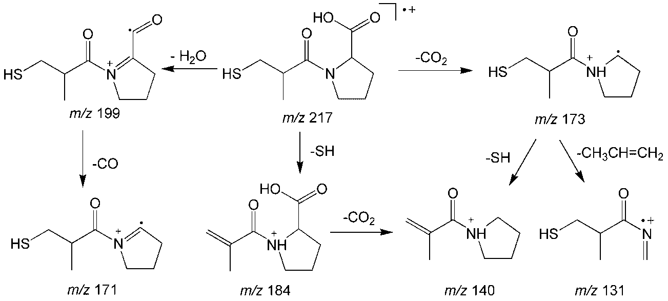

 Scheme 1. Proposed fragmentation pathways for the captopril cation.

 Scheme 2. Proposed fragmentation pathways for the fudosteine cation.
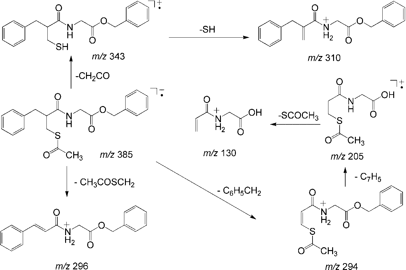
 Scheme 3. Proposed fragmentation pathways for the racecadotril cation.